iThera Medical’s optoacoustic imaging (OAI) systems are based on the photoacoustic effect. In 1881, Alexander Graham Bell discovered that light energy absorbed by a material results in an acoustic signal. While Alexander Graham Bell used sunlight as incident light and a hearing tube as acoustic detector, modern optoacoustic imaging systems use high-energy pulsed lasers and highly sensitive broadband ultrasound detectors. By exciting tissue with a laser pulse and detecting the signals generated by the conversion of light energy into sound waves, optical absorption in tissue can be detected and visualized.
Leveraging the photoacoustic effect, OAI has the unique capability of visualizing optical contrast at high resolution (down to 10 µm) in deep tissue (up to 3 cm) and displaying the image in real time. The frame rate is dependent on the pulse repetition rate of the light source and can reach up to hundreds of Hertz.
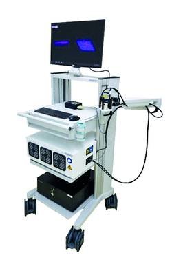
iThera Medical is the only company offering OAI systems in the mesoscopic regime – achieving 2-3mm depth penetration at a resolution of down to 20µm. Raster-scanning optoacoustic mesoscopy (RSOM) has so far been explored in a variety of tumor-associated preclinical studies, using the RSOM Explorer P50 to look at angiogenesis and changes in tumor vasculature under treatment. For clinical imaging, the RSOM Explorer C50 has been used for clinical trials in skin cancer and a wide range of skin diseases associated with inflammation.
Due to its favorable characteristics in terms of spatiotemporal resolution, penetration depth, sensitivity and specificity, OAI can be used for a multitude of applications, for both preclinical and clinical research:
- Tumor characterization
- Functional brain imaging
- Assessment of organ function
- Nanoparticle tracking
- Crohn’s disease imaging
- Breast cancer imaging
- Melanoma detection
- Other preclinical and clinical applications
Currently there is the only one RSOM iThera Medical system for preclinical research in Russia. System was delivered and installed by Spektronika LLC and is already being successfully used by Biophotonics laboratory scientists in Skoltech (Skolkovo institute of science and technology).